
Article · 10 November 2026
Is open data ready to use?
21 medical datasets, 13 open models
I am a physician. I worked in emergency departments for three years and I am now a pediatric surgery resident. I want AI to help us make a diagnosis faster, lose no time in emergencies, and give every patient the same chance. That is why I started training models myself, and why I started with open data.
In the past weeks I prepared 21 open medical datasets and published them on EVREN, Türkiye's national AI platform, with 13 open baseline models. The 13 models took 28 training runs. With experiments and private work, I trained more than 100 models. The datasets cover surgical video (pancreatic veins, gastric vessels, gastrectomy, rectal anatomy), endoscopy and bronchoscopy, fetal and pelvic floor ultrasound, ECGs, urine and semen microscopy, coronary angiography and wrist X-rays. The collection: evren.ssyz.org.tr/collections/01a1b752-1527-7231-8d73-3e4174e74810.
The vein model
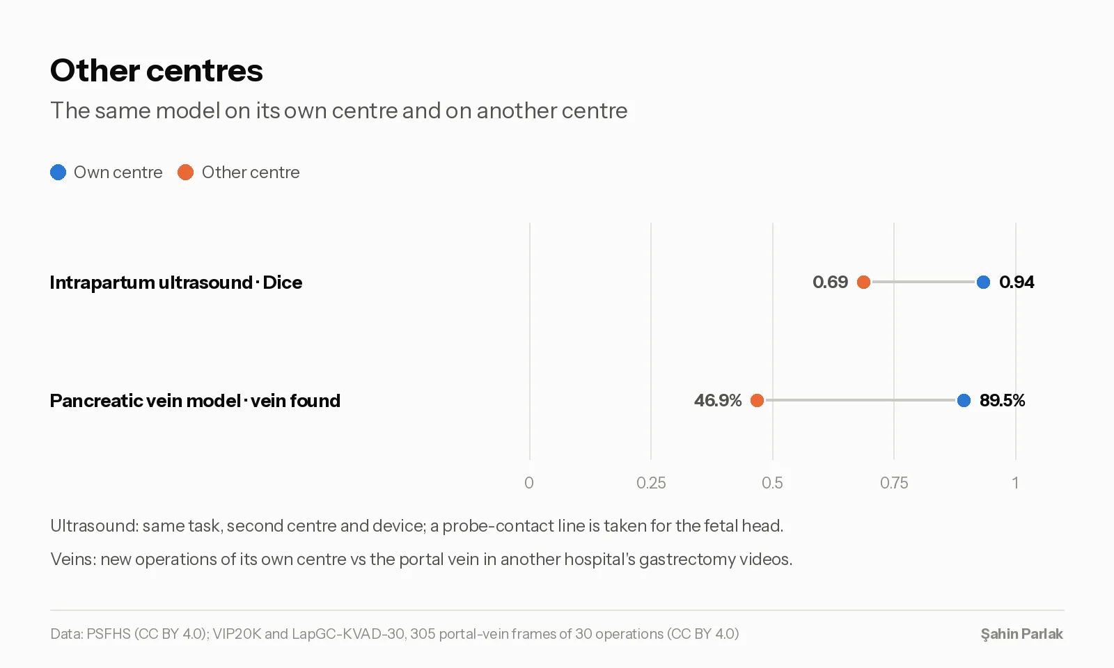
One of the models finds the large veins in laparoscopic pancreatic surgery: the superior mesenteric, portal and splenic veins. I trained it on VIP20K, a dataset from Peking Union Medical College Hospital. On four operations it had not seen, it found the vein in 89.5% of frames.
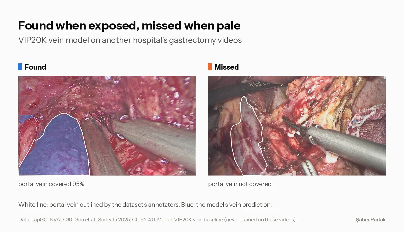
Then I tested it on gastrectomy videos from another hospital (LapGC-KVAD-30, 30 operations). Only the portal vein is labelled there. The model found it in 47% of frames. When the vein was well exposed, bulging and dark, the model covered almost all of it. When it was flat, pale or under a thin layer of tissue, the model did not see it.
A surgeon knows both views of the portal vein. The model knew only one, because it had seen only one. When I saw it miss the vein, I understood that the question is not which system is best. It is what data we train it on.
So, is open data ready to use? After 21 datasets, my answer is: usually yes, but not as you download it. These are the checks I made.
Licenses
Many medical datasets exist in several places. There is the authors' own record (Zenodo, figshare, PhysioNet, a journal), and there are copies on Kaggle, Roboflow, Hugging Face or Mendeley. The copies often show a license that the original does not give.
Some examples from chest X-ray sets. A Mendeley copy of the Shenzhen and Montgomery tuberculosis images says CC BY, but the original terms say the data must not be shared outside the research group. The pediatric pneumonia set of Kermany et al. says “for use in research only” in version 3, but not in version 2. The LNDb lung CT record says CC BY in one field and CC BY-NC-ND in its description. Several “laparoscopy” sets marked CC BY or MIT were copies of non-commercial data.
The paper and the data can also have different licenses. LapGC-KVAD-30 data is CC BY 4.0, but its article is CC BY-NC-ND. I used the data and none of the article's figures.
So I read every license at the original source and write down where and when. I upload nothing before that. Most of the well-known chest X-ray collections do not allow open sharing.
Duplicates and leaks
I split every dataset by patient, by operation for surgical video, or by group where there are no patient IDs, so that images of one person are never in both training and test. This is necessary, but it is not always enough. The same data sometimes appears under two names.
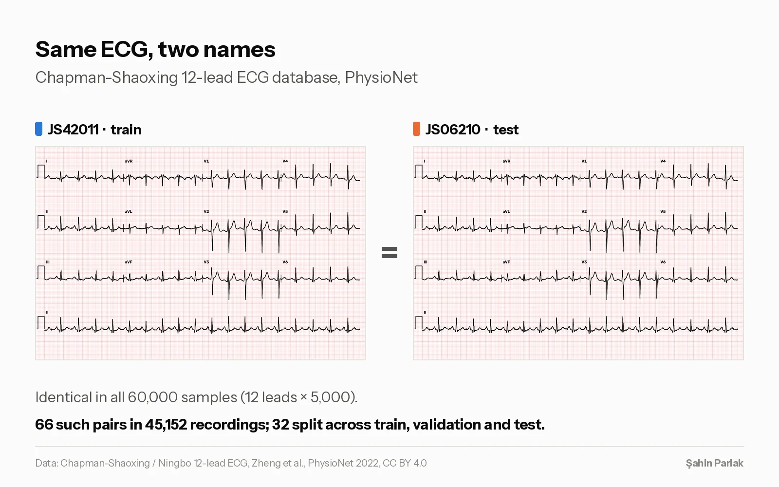
The clearest case was an ECG database. Chapman-Shaoxing has 45,152 recordings. 66 of them appear twice, under different record names, and the signals are identical. After my split, 32 of these pairs were on both sides: one copy in training, one in validation or test. I did not catch this myself. EVREN checks every file on import and warned me about identical files. I compared the raw signals and removed one copy of each pair.
There were others:
- ECG Images of Cardiac Patients: 928 files, but only 491 different images.
- PFUS1 (pelvic floor ultrasound): 6 duplicated video folders, 946 frames.
- FOCUS (fetal heart): one image in both the official training and test sets. MODUS (urine sediment): five such pairs.
- HyperKvasir (endoscopy): the official folds split 20 of 42 near-identical image pairs.
Exact copies are easy to find with file hashes. Near-copies, like the same polyp filmed a few seconds apart, are harder. I used image similarity and then looked at the unclear pairs myself. In the HyperKvasir polyps I checked 416 pairs and grouped 30. This has limits: fetal ultrasound images of different mothers can look so alike that similarity cannot separate them.
In ARCADE, PSFHS and PFUS1, some identical images had different outlines. This shows how precise the labels really are.
Simple baselines
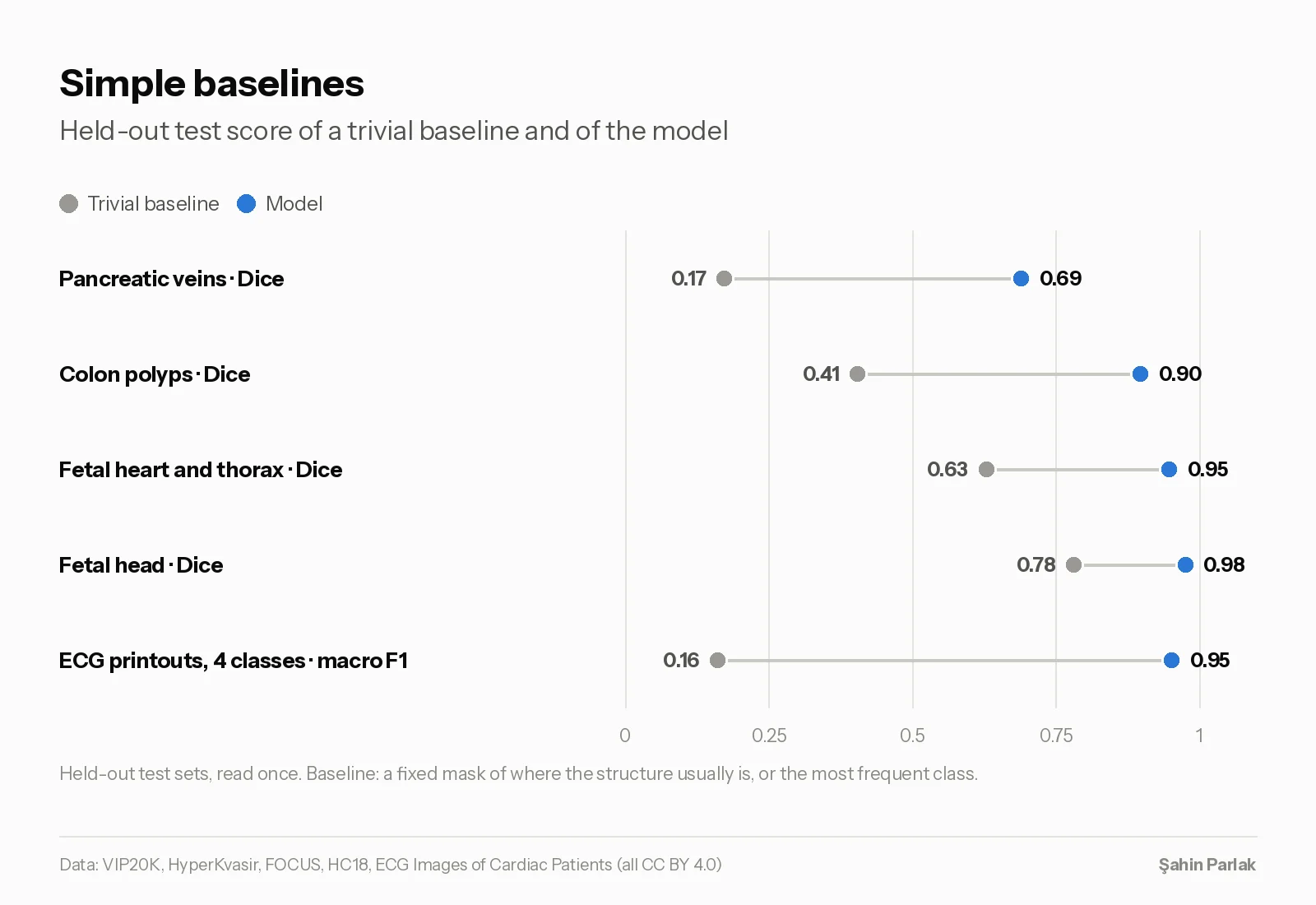
In HC18, a Dice score of 0.78 for the fetal head sounds good. But a fixed oval in the middle of the image gets 0.78 without looking at the image. The model gets 0.976. More important for the clinic, it measures head circumference with a mean error of 2.32 mm. The fixed oval is off by 46.2 mm.
For pancreatic veins, a fixed mask of where veins usually are gets Dice 0.17, and the model 0.69. For colon polyps it is 0.41 against 0.90. The fixed mask can even look better on the wrong measure: on the other hospital's videos it “finds” the portal vein more often than the model (71% against 47%), because it covers 23% of every frame.
I have run this baseline for five of the 13 models so far. The other eight still need it.
Two runs per model
I trained every published model twice with the same settings and a different random seed. In most datasets the two runs ended within 0.01 of each other. Single classes moved by up to 0.19. In 3 of 11 cases, the run I chose on validation was a little worse on the test set than the other run. I fix the rule for choosing before I look at the test, and I look at the test only once.
Other centres
Validation and test results were usually close. VISEM (sperm videos) was an exception: with only three men per split, which men were in a split changed the result more than anything I did.
The big drops came from other centres, like the portal vein. PSFHS (intrapartum ultrasound) has images from two centres and two devices. The model got Dice 0.94 on images from its own centre and 0.69 on the second device. There it took a bright probe-contact line for the fetal head. You do not see this if you test only on the training centre.
Reading training pages
Some points to keep in mind on EVREN and on most platforms. The main score on a model page is a box metric, even for segmentation models, so a higher score does not always mean better masks. The validation loss shown does not include the mask term, but the training loss does. The saved “best” model is the best epoch on validation, and it can be a very early one. I evaluate every model again against the original masks before I report a number.
My request to readers
Most of these datasets are excellent work by their authors. All 21 are now open, with licenses checked at the source, splits by patient or group, and every change documented. 13 of them also have a baseline model. None of the models is a clinical tool.
If you use them, or any open medical data, please do not copy and paste it into your project. Pass it through a controlled filter first, adapt it to your own field, and make something that helps people.
Collection: evren.ssyz.org.tr/collections/01a1b752-1527-7231-8d73-3e4174e74810 · Scripts: github.com/sahinparlak/open-medical-baselines